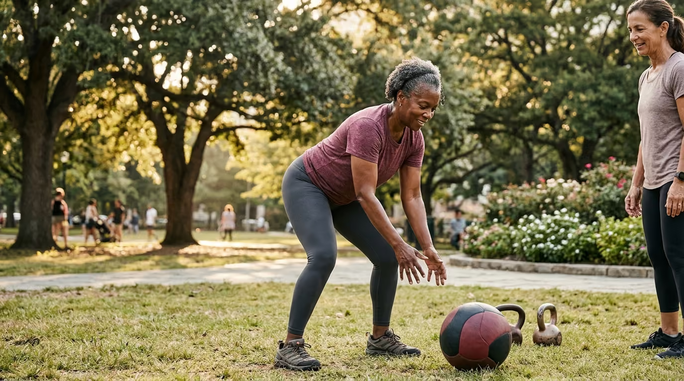

Stronger bones and reduced fracture risk after menopause come from combining progressive resistance, weight-bearing impact, balance drills, and posture training.





Exercise for bone strength after menopause is not a simple recommendation for light daily walking or gentle toning. It is a targeted combination of progressive muscle loading, controlled impact forces, postural training, and dynamic balance work designed to reinforce the physical skeleton. Bone health is distinct from cardiovascular fitness. While aerobic activity supports heart health and metabolic capacity, bone tissue requires specific mechanical stress to stimulate remodeling and maintain structural integrity.
This resource provides a complete, research-backed guide to training your skeletal system through midlife and beyond. It explains how mechanical forces act on bone tissue, how exercise modalities vary in their effects, and how to build a weekly program tailored to your density measurements and fracture history. Understanding these physical principles helps women maintain capability and confidence while protecting long-term mobility. Explore our strength and body composition guide for further insights into physical function across midlife.
The biological transition into menopause introduces significant shifts in skeletal remodeling. Bone tissue is constantly breaking down and rebuilding through a continuous cellular process. Specialised cells called osteoclasts remove older bone tissue, while osteoblasts form new bone matrix. Estrogen plays a protective role in this cycle by regulating osteoclast activity and preventing excessive resorption.
When estrogen levels fluctuate and permanently decline during perimenopause and menopause, this balance shifts. Resorption begins to outpace new bone formation. Clinical data indicates that bone loss often begins two to three years before the final menstrual period. During this transition, women can lose approximately 1.5% of their lumbar spine bone mineral density each year.
This loss often accelerates during the first two years following the final menstrual period. Rates of bone loss during this window can reach up to 2.5% annually before gradually slowing to a lower baseline rate. Because these figures represent population averages, individual experiences vary based on genetics, baseline peak bone mass, body composition, and physical activity history.
To evaluate these changes, clinicians rely on dual-energy X-ray absorptiometry, commonly known as a DXA scan. A DXA scan measures bone mineral density at critical anatomical sites, primarily the lumbar spine, femoral neck, and total hip. Results are reported as a T-score, which compares a person's density to the young-adult reference average:
A T-score provides useful diagnostic information, but it does not tell the whole story of skeletal health. Two individuals with an identical T-score of −2.2 can have vastly different physical capabilities, fall risks, and muscle strength. Exercise planning must look beyond the scan to evaluate movement skill, balance, posture, and prior injuries. Learn more about managing hormonal transitions in our section on perimenopause and menopause articles.
Credible research confirms that physical exercise is a fundamental component of maintaining postmenopausal bone health and reducing fracture risk. Systematic reviews show that dynamic resistance training and weight-bearing exercises can reduce bone loss at the spine and hip compared to inactive control groups. Clinical guidelines globally recommend regular muscle-strengthening and weight-bearing activity as standard care for midlife women.
However, the evidence also reveals clear nuances regarding what exercise can and cannot achieve:
Meta-analyses of exercise trials in postmenopausal women frequently demonstrate statistically significant improvements in bone mineral density at the lumbar spine and femoral neck. However, the absolute density gains are typically modest, often ranging between 1% and 3% over a training period. Exercise rarely restores low bone density back to young-adult levels on a DXA scan.
Bone strength involves more than mineral density alone. It includes bone size, shape, cortical thickness, and internal architecture. Dynamic mechanical loading alters bone geometry and redistributes mass to areas experiencing higher stress. These structural improvements enhance mechanical capacity, even when a standard two-dimensional DXA scan shows minimal change in numerical density.
A major mechanism by which exercise prevents fractures is by stopping falls from happening in the first place. Most osteoporotic fractures, particularly of the hip and wrist, occur as a direct result of a fall from standing height. Multicomponent exercise programs that combine muscle strengthening with challenging balance drills significantly reduce fall rates in older adults.
The total body of evidence shows that exercise protects the skeleton through a combined mechanism. It provides local mechanical strain to slow bone density loss while simultaneously improving muscle strength, reaction time, gait stability, and falling resistance.
An effective exercise framework for postmenopausal bone health relies on four complementary pillars. Relying on a single modality, such as daily walking or light stretching, omits key physical adaptations necessary for full skeletal support.
Progressive resistance training forces muscles to pull against external loads or body mass, generating tensile and compressive forces on attached bones. To stimulate bone formation, resistance training must apply mechanical strain that exceeds routine daily tasks.
Targeted muscle groups should include major structural attachments:
Training parameters recommend performing resistance exercises at least twice weekly. Programs typically feature six to eight exercises targeting major muscle groups, executing two to three sets of eight to twelve repetitions at a moderate-to-hard rating of perceived exertion.
Weight-bearing activity requires the body to support its own mass against gravity. Impact loading adds rapid acceleration and deceleration forces, which generate ground reaction forces that travel through the lower limbs and spine.
Impact loading exists along a dynamic continuum:
Dynamic, high-rate forces stimulate osteoblast activity efficiently. However, impact must be selected based on baseline joint health, current bone density, landing mechanics, and fall risk.
Balance training trains the central nervous system and muscular system to maintain control over the body's center of mass. Effective fall-prevention routines go beyond resting on one leg for a few seconds; they involve active, challenging movement patterns.
Key elements of dynamic balance work include:
Guidelines recommend practicing balance exercises for 15 to 20 minutes daily, aiming for roughly two hours of dedicated balance work each week.
Spinal health requires specific attention to posture, trunk endurance, and safe movement mechanics. Vertebral compression fractures often occur during unassisted forward bending combined with heavy twisting or lifting.
Postural training focuses on strengthening the spinal extensor muscles and deep trunk stabilizers. Improving back extensor endurance helps maintain a neutral spine during everyday activities like lifting items from the floor, carrying groceries, or bending over a garden bed.
Systematic progression is essential to build skeletal strength without overstressing joint tissue or triggering injury. Applying the principle of progressive overload means gradually increasing mechanical demands as your muscles and skeletal tissue adapt.
Beginners should establish proper movement mechanics using body weight, light dumbbells, or resistance machines before adding heavy external loads. A practical sequence for progressive loading involves:
Impact training should be introduced in a staged manner. Jumping immediately into high-impact workouts without preparation can cause joint pain or soft-tissue injury.
Use this step-by-step impact ladder to guide progression:
Progress up the ladder only after completing four to eight weeks of consistent resistance training. Progression should only occur if you remain completely pain-free during and after activity.
Exercise is beneficial across all bone density levels, but safety guidelines vary significantly based on your diagnostic history and fracture background.
Individuals with osteopenia generally have low structural restrictions unless they have suffered a prior fragility fracture or have high fall risks.
Individuals diagnosed with osteoporosis without prior fractures can lift challenging weights and participate in dynamic exercise when performed with correct mechanics.
A history of spinal fracture requires specific adaptations to protect the spinal column from high compressive and shearing forces.
If you have sustained a hip fracture or experience frequent balance loss, fall prevention becomes the central focus.
Exercise does not occur in biological isolation. Mechanical stress provides the signal for bone growth, but the body requires adequate nutritional resources and medical oversight to complete the remodeling process.
Calcium provides the physical building blocks for bone matrix, while vitamin D is required for intestinal calcium absorption and muscular function.
Muscle mass and bone density are closely linked. Skeletal muscle applies direct mechanical pull on bone, and lean mass contributes to overall force output. Maintaining muscle mass through adequate dietary protein intake and resistance training helps protect the skeleton during age-related hormonal shifts. Read our overview of midlife health on our about Refemina page.
Prolonged sitting promotes bone resorption and reduces muscle activation. Interjecting long sedentary stretches with short standing breaks, stair walks, or brief movement sessions supports bone health alongside structured exercise routines.
Exercise is an essential component of bone health, but it does not replace medical treatment when clinical intervention is required. Individuals with severe osteoporosis or high short-term fracture risks may require prescription bone-active medications alongside an active exercise program.
While exercise research offers clear positive evidence, understanding its limitations prevents unrealistic expectations and ensures safe application.
Research literature includes a wide variety of exercise types, durations, and intensity levels. Some clinical trials utilize high-intensity resistance training under direct supervision, while others rely on home-based low-impact routines. As a result, pooling meta-analysis data can mask which specific protocols yield the greatest adaptations.
Standard two-dimensional DXA scans measure planar bone mineral density ($g/cm^2$). They cannot measure three-dimensional bone geometry, trabecular microarchitecture, or bone quality changes. An individual may build stronger bone architecture through exercise without seeing dramatic changes in raw DXA numbers.
Most research trials investigating high-load resistance or jump training deliberately exclude individuals with severe osteoporosis, acute back pain, or recent vertebral fractures. Data supporting high-intensity training programs cannot be automatically applied to high-risk individuals without clinical modifications.
Bone remodeling is a slow biological process. Most published exercise studies last between six months and two years. Understanding the full lifetime impacts of exercise on age-related fracture rates requires long-term observational monitoring.
These practical templates demonstrate how to organize resistance, impact, balance, and weight-bearing activities into a structured weekly routine.
Consulting a medical doctor, physical therapist, or certified exercise specialist is necessary when designing an exercise plan in the presence of specific health factors.
A specialized physical assessment should examine:
Replacing general prohibitions with specific movement cues builds self-confidence and ensures physical activity remains safe and effective. Access additional evidence-based planning tools in our menopause and midlife resources section.
Swimming and cycling offer cardiovascular benefits and preserve muscle mass, but they are non-weight-bearing activities. Because water supports body weight and cycling pedals smooth out impact, these exercises generate minimal ground reaction forces or skeletal strain. They should be supplemented with progressive resistance training and weight-bearing land exercises to support bone density.
Yoga and Pilates build core endurance and flexibility, but certain postures require modification for people with osteoporosis. Exercises involving deep forward spinal flexion, dynamic toe-touching, or weighted spinal rotation should be modified. Focus on postures that emphasize neutral spine alignment, back extensor strength, and hip mobility instead.
To stimulate bone modeling, resistance exercises should feel challenging by the final repetitions of a set while maintaining safe technique. On a 1-to-10 rating scale of perceived exertion, the effort should land between 7 and 8 (hard to very hard). If you can complete 15 to 20 repetitions without muscular fatigue, the load is likely too light to maximize skeletal stress.
Bone tissue adapts slowly. Measurable changes in mineral density on a DXA scan typically take 12 to 24 months of consistent training to appear. However, improvements in muscle strength, dynamic balance, gait speed, and postural control happen much faster, often within 8 to 12 weeks of starting a program.
Postmenopausal bone exercise should be progressive without being reckless, protective without inducing fear, and individualized rather than defined by a single T-score. The goal is to build a resilient skeletal and muscular system that allows you to produce force, manage balance, handle routine physical loads, and remain active over your lifetime.

Follow Refemina for research-led resources on hormonal change, strength, metabolism, nutrition, skin, intimate wellbeing and confidence through midlife.


Keep learning about the changes that shape energy, strength, confidence and wellbeing through midlife.
Explore Resources