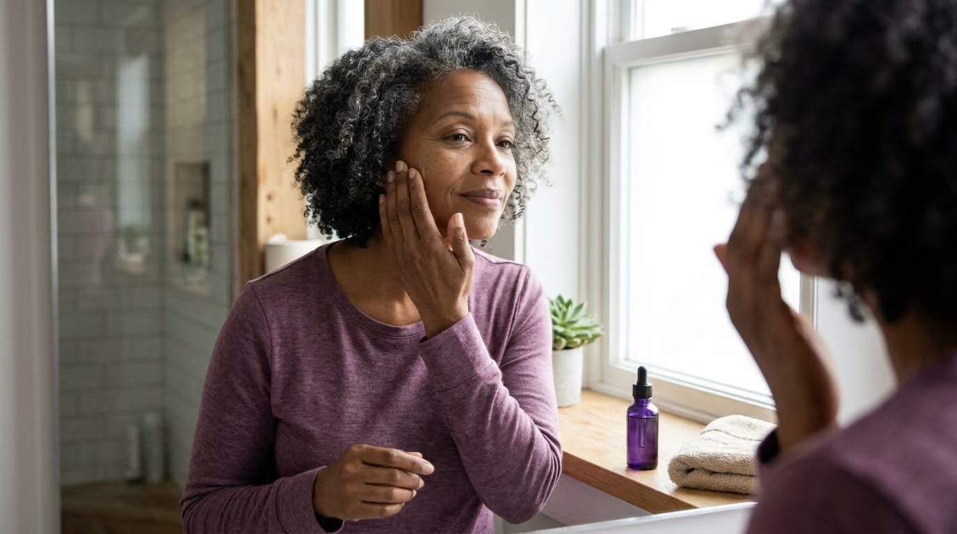

Midlife skin changes involve far more than surface wrinkles, as shifting hormones simultaneously drive dermal thinning, stubborn jawline breakouts, dryness, and vascular flushing.





Menopause skin change is not a single clinical condition, nor is it an inevitable cosmetic crisis. It is the complex intersection of endocrine shifts, chronological aging, cumulative solar radiation, genetic factors, daily personal care routines, and underlying health.
When ovarian hormone production changes during perimenopause and postmenopause, the biological signals that govern skin thickness, lipid production, repair capacity, and water retention shift alongside it. At the same time, decades of environmental exposure and natural cellular aging continue their steady progression. Understanding these changes requires separating biological realities from marketing claims, allowing you to care for your skin with clarity rather than frustration.
This resource breaks down the mechanisms behind midlife skin changes, examines the evidence surrounding treatment options, and offers a practical framework for supporting comfort, resilience, and skin health across the menopause transition.
Research demonstrates that skin is a major hormone-responsive organ. Receptors for estrogen, progesterone, and androgens are located throughout epidermal keratinocytes, dermal fibroblasts, sebaceous glands, hair follicles, and blood vessel walls. When hormone concentrations change, these cells alter their structural output and maintenance cycles.
Current evidence separates skin aging into two distinct processes that interact continuously:
Clinical research shows that systemic estrogen loss reduces dermal collagen content, thins the epidermal layer, decreases skin hydration, and alters the mechanical properties of elasticity. A landmark analysis found that skin collagen decreases significantly in the years following menopause, with an estimated loss of nearly thirty percent during the first five postmenopausal years, followed by an average annual decline of about 2.1 percent over the subsequent fifteen years. These figures represent study averages rather than a precise script for every woman, yet they explain why structural skin changes often seem to accelerate in midlife.
At the same time, dermatological studies emphasize that changing hormone levels do not act in isolation. Everyday cleansing habits, ambient humidity, medications, nutritional status, and systemic health dictate how these physiological shifts appear on your face and body. You can explore broader perspectives on midlife physiology in our menopause and hormonal change resources.
The menopausal transition involves distinct phases. Perimenopause introduces wide fluctuations in estrogen and progesterone, while postmenopause establishes a permanent low-estrogen state. These hormonal movements alter multiple layers of the skin architecture.
Dermal fibroblasts rely partly on estrogen signaling to synthesize type I and type III collagen, the primary scaffolding proteins responsible for firmness and tensile strength. Estrogen also supports the production of glycosaminoglycans, including hyaluronic acid, which reside in the extracellular matrix and bind water molecules to create tissue fullness.
With declining estrogen, fibroblast activity slows down while enzymes that break down matrix proteins, known as matrix metalloproteinases, remain active. This imbalance leads to dermal thinning and reduced elasticity. The skin becomes more prone to creping, laxity, and fine surface lines. Improving surface hydration can soften the visual appearance of these fine lines, but topical moisturizers cannot rebuild degraded dermal architecture.
The epidermis serves as the frontline shield against environmental stressors, pathogens, and excessive moisture loss. Estrogen supports normal keratinocyte proliferation and the organization of the stratum corneum, the outermost layer of dead skin cells embedded in a specialized lipid matrix.
During the menopause transition, epidermal renewal slows. The production of essential barrier lipids, including ceramides, free fatty acids, and cholesterol, declines. This structural disruption weakens the skin barrier, leading to increased transepidermal water loss. The result is a skin surface that struggles to retain moisture, feels chronically tight, and reacts rapidly to common topical ingredients.
Estrogen supports capillary blood flow and vascular endothelial health throughout the body. In the dermis, reduced estrogen levels lead to decreased capillary density and diminished microcirculation.
This reduction in blood supply limits the delivery of oxygen and micronutrients to the upper layers of the skin. It also slows cellular repair mechanisms. Cuts, scrapes, and blemishes may take longer to resolve, and skin can take on a more translucent or pale appearance as vascular support changes.
Dry skin, clinically termed xerosis, is one of the most frequent dermatological complaints reported in midlife. A clinical study of 150 postmenopausal women evaluated in a specialized setting identified xerosis in 78 percent of participants. While this figure comes from a clinical sample seeking care and should not be viewed as an absolute population statistic, it reflects the widespread nature of the issue.
Xerosis is not merely a lack of water. It is a failure of the lipid barrier that holds water inside the tissue. When ceramide and sebum production drop, microscopic gaps appear between surface skin cells. Water evaporates rapidly into the surrounding air, leaving the skin rough, flaky, and prone to fine scaling.
This barrier deficit creates a common cycle of irritation:
Pruritus, or persistent itching, often accompanies severe xerosis. Dry, compromised skin fires sensory nerve endings more easily, sending itch signals to the brain even without an obvious rash.
However, persistent or severe itching should not automatically be dismissed as normal menopausal dryness. Generalized pruritus can occasionally stem from thyroid dysfunction, iron deficiency, renal or hepatic disease, or underlying contact dermatitis. If thorough moisturization and gentle skin care do not relieve persistent itching, a thorough medical review is warranted.
One of the most frustrating experiences in midlife is developing inflammatory acne while simultaneously struggling with dry, flaking skin. Many women assume acne is strictly a teenage concern linked to oily skin, yet midlife breakouts follow a distinct hormonal mechanism.
Acne in perimenopause and menopause rarely stems from an absolute surge in testosterone. Instead, it occurs because ovarian production of estrogen and progesterone declines more rapidly than adrenal and ovarian androgen production.
This creates an environment of relative androgen predominance. Androgen receptors in the sebaceous glands remain active, stimulating localized sebum production and altering the composition of facial oils. Combined with slower cellular turnover in the hair follicle lining, pores become clogged with dead skin cells and sticky sebum, creating an ideal setting for acne-causing bacteria.
Hormone-responsive adult acne frequently concentrates along the lower third of the face, appearing along the jawline, chin, and upper neck. These lesions often present as tender, deeper inflammatory papules or nodules rather than superficial blackheads.
Treating this form of acne requires care. Traditional acne regimens designed for adolescent skin rely on harsh astringents, high-strength salicylic acid, and aggressive drying agents. When applied to menopausal skin with a compromised lipid barrier, these products strip surface moisture without resolving the underlying hormonal trigger. The result is inflamed, painful acne sitting on top of raw, peeling skin.
Consider a realistic clinical example. A 52-year-old woman notices cystic blemishes along her chin while her cheeks and temples feel tight and rough. In response, she adopts a foaming clarifying wash, an alcohol toner, and daily spot treatments. Within two weeks, her jawline remains broken out, but her face now burns upon applying even basic lotion.
The evidence-aligned solution is not to attack the acne with stronger drying products. Instead, the approach centers on repairing the general lipid barrier while using targeted, non-drying acne therapies only on the breakout zones. Balancing these concerns requires patience and a tailored routine, which you can read more about across our skin, hair, and aesthetic aging resources.
Many women notice that products they used comfortably for decades suddenly trigger redness, warmth, or stinging. Midlife skin reactivity involves several overlapping vascular and neurological factors.
Vasomotor symptoms, commonly known as hot flashes, stem from temperature-regulation shifts in the brain, resulting in transient facial and upper-body flushing. However, true rosacea is an independent chronic inflammatory disorder characterized by persistent central facial erythema, visible telangiectasias (broken capillaries), and inflammatory bumps.
While hot flashes and rosacea are biologically distinct, the repeated vascular dilation from hot flashes can aggravate underlying rosacea. Differentiating between the two is vital:
Managing facial reactivity begins with identifying personal triggers. Heat, sun exposure, spicy meals, red wine, emotional stress, and intense exercise can provoke cutaneous flushing. While eliminating triggers does not cure rosacea, tracking them helps minimize severe inflammatory flares.
Hyperpigmentation changes become more prominent during midlife, reflecting both past environmental exposure and shifting hormonal signals. Understanding the nature of different pigmentary patterns prevents unnecessary treatments that can cause further discoloration.
Uneven skin tone generally falls into one of three categories:
When dark patches appear, the instinct is often to use harsh bleaching creams, aggressive physical scrubs, or unmonitored chemical peels. In midlife skin, an already vulnerable barrier responds to harsh treatments with inflammation.
This inflammation frequently stimulates melanocytes to deposit more pigment, making melasma or dark marks darker than before. Evidence-based pigment management relies first on broad-spectrum ultraviolet protection, followed by gentle, consistent tyrosinase inhibitors prescribed or recommended by a qualified clinician.
The relationship between menopausal hormone therapy and skin quality has been investigated in multiple clinical trials, yet the scientific findings require nuanced interpretation.
Systemic hormone replacement therapy provides proven relief for vasomotor symptoms and helps prevent bone loss. Researchers have also documented measurable improvements in skin parameters among women using systemic estrogen therapy.
In a randomized, double-blind, placebo-controlled trial evaluating sixty postmenopausal French-Canadian nuns over twelve months, systemic conjugated equine estrogen increased total skin thickness by 11.5 percent and dermal thickness by 33 percent compared to the control group. Other studies summarized in systematic reviews report improvements in dermal collagen density, surface hydration, and measured elasticity among women using systemic estrogen.
However, clinical organizations such as the American College of Obstetricians and Gynecologists and The Menopause Raleigh Society do not recommend systemic hormone therapy solely for cosmetic purposes or anti-aging indications. Systemic hormone therapy carries specific clinical considerations, indications, and contraindications that must be evaluated with a physician based on overall health, rather than skin appearance alone. Any dermatological benefits should be viewed as a secondary outcome of medically indicated treatment.
Topical estrogen creams applied directly to facial skin have also been studied. Systematic reviews indicate that low-concentration topical estrogen can increase dermal collagen concentration, improve hydration, and reduce the appearance of fine wrinkling in small clinical cohorts.
However, the evidence for topical estrogen remains limited. Formulations, concentrations, application frequencies, and study endpoints vary widely across published trials. Furthermore, topical estrogen can undergo systemic absorption, which raises unanswered safety questions for individuals with contraindications to hormone therapy. At present, topical estrogens are not widely approved by major regulatory bodies as standard cosmetic therapies for aging skin.
Collagen powders, drinks, and botanical supplements are heavily promoted for menopausal skin, yet high-quality independent evidence remains modest. While hydrolyzed collagen peptides show some potential in small industry-sponsored trials to support hydration and elasticity, they are broken down into basic amino acids in the digestive tract before absorption.
They do not travel directly to facial skin to assemble new collagen fibers. A balanced diet containing adequate dietary protein, vitamin C, zinc, and healthy fats provides the foundational building blocks for cellular repair without relying on expensive niche formulations.
Caring for skin in perimenopause and postmenopause does not require an elaborate twelve-step routine. Dermatological consensus highlights that simplifying your approach, respecting the moisture barrier, and protecting against ultraviolet radiation deliver the most reliable results.
Cleansing should remove dirt, pollutants, and sunscreen without stripping the delicate lipid film from the stratum corneum.
A functional moisturizer for mature skin should contain a balance of three distinct ingredient categories:
Apply your moisturizer immediately after washing while the skin is still damp to seal in surface water.
If you wish to address breakouts, uneven pigmentation, or cellular renewal, introduce active ingredients slowly and one at a time.
Ultraviolet protection remains the single most effective intervention for preventing premature collagen breakdown, solar elastosis, hyperpigmentation, and skin cancers.
Use a broad-spectrum sunscreen with an SPF of 30 or higher every single morning, regardless of the weather. Mineral options containing zinc oxide or titanium dioxide are often particularly well tolerated by reactive, rosacea-prone, or sensitive skin. Sunscreen will not erase existing structural changes, but it halts ongoing UV-mediated collagen loss and prevents melasma from darkening.
While many midlife skin changes reflect normal biological shifts, several clinical signs warrant evaluation by a dermatologist, primary care clinician, or gynecologist.
Seek prompt clinical evaluation if you experience any of the following:
A qualified clinician can establish an accurate diagnosis, rule out systemic conditions, and prescribe targeted medical therapies when over-the-counter measures prove inadequate. You can learn more about finding evidence-based guidance in our educational resource library.
Drinking adequate water is important for overall health, but systemic hydration alone cannot resolve xerosis caused by an impaired lipid barrier. If the skin lacks ceramides and natural moisturizing factors, water simply evaporates through the stratum corneum. Topical moisturizers containing humectants, emollients, and occlusives are necessary to seal hydration within the tissue.
Estrogen decline leads to thinning of both the dermis and the protective subcutaneous fat layer, while microvascular fragility increases. Without a thick collagen cushion surrounding dermal blood vessels, minor bumps that once left no mark can cause small capillaries to rupture, resulting in visible bruising. If bruising occurs spontaneously without trauma, spreads rapidly, or is accompanied by other bleeding symptoms, consult a physician.
You do not necessarily need to discontinue retinoids permanently, but you should adjust how you use them. Reduce your application frequency to once or twice a week, apply a generous layer of barrier-supportive moisturizer first, and let it absorb before applying a pea-sized amount of retinoid. If burning, stinging, or redness persists, pause the active until your barrier fully recovers.
Not necessarily. In most menopausal women experiencing breakouts, total testosterone levels are normal or even declining. The breakouts occur because estrogen and progesterone levels have dropped more steeply, leaving a higher ratio of androgens relative to estrogens. This relative imbalance is enough to stimulate sebaceous glands and trigger lower-face acne.
Understanding the biology of your skin during perimenopause and beyond allows you to replace aggressive anti-aging routines with supportive, restorative care. By prioritizing your moisture barrier, protecting against solar exposure, and treating specific concerns with gentle precision, you can maintain skin comfort, resilience, and health throughout midlife.

Follow Refemina for research-led resources on hormonal change, strength, metabolism, nutrition, skin, intimate wellbeing and confidence through midlife.


Keep learning about the changes that shape energy, strength, confidence and wellbeing through midlife.
Explore Resources